Axiora Medical
Mandibular reconstruction is not simply a matter of selecting the strongest metal plate. It is a clinical decision involving bone loss, occlusion, soft-tissue coverage, infection risk, and the patient’s long-term function. The International Agency for Research on Cancer reported 389,846 new lip and oral cavity cancer cases worldwide in 2022. Many advanced tumors require segmental mandibulectomy, creating complex structural demands for reconstruction.
Mandibular Reconstruction Plates must restore continuity while supporting chewing, speech, facial symmetry, and healing. Surgeons typically review defect length, remaining bone quality, dental status, radiation exposure, and the availability of vascularized tissue. Plate geometry also matters. A reconstruction plate crossing the mandibular angle experiences different mechanical forces from one supporting the anterior arch. Screw selection, spacing, contouring accuracy, and load distribution can influence stability.
Small details matter.
AO CMF surgical principles emphasize accurate reduction, stable fixation, and protection of soft tissues. The U.S. Food and Drug Administration also expects manufacturers to demonstrate device safety, performance, and biocompatibility through appropriate evidence. However, published complication rates vary between studies because patient conditions and surgical techniques differ. This makes direct comparisons unreliable.
There is no universal best plate.
A practical selection process should balance rigidity, contourability, screw options, anatomical fit, and the possibility of secondary reconstruction. Clinical teams should also consider manufacturer documentation, traceability, sterilization requirements, and postoperative imaging. An experienced surgeon may still revise the plan intraoperatively when bone quality or flap coverage differs from preoperative imaging. That uncertainty deserves attention. The ideal choice is not necessarily the thickest plate, but the one that matches the defect, tissue environment, and rehabilitation goals.
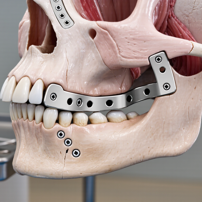
Mandibular reconstruction plates restore continuity after tumor removal, trauma, or severe bone loss. Their main function is mechanical support. They also help maintain facial width, bite alignment, and mandibular contour during healing. Plate selection depends on defect length, bone quality, occlusion, and soft-tissue condition.
Load-bearing plates carry most chewing forces when bone cannot provide stable support. They are often considered for segmental defects or weakened residual bone. Load-sharing plates work differently. They depend on contact between bone segments and usually suit more stable fractures.
Locking systems can maintain screw-plate stability when bone is thin or uneven. Nonlocking systems may be useful when close plate-to-bone contact is achievable.
Shape matters too. Straight plates may fit simple defects, while curved or angled designs can follow the mandibular border more naturally.
A patient-specific plate may improve contour accuracy, but it still requires careful imaging and surgical judgment. Surgeons should check screw length, nerve pathways, dental occlusion, and soft-tissue coverage. A plate that looks ideal on a scan may feel wrong in the operating field. That gap deserves attention.
Current evidence supports sound planning, but no plate type works perfectly for every patient. Parallel fixation, adequate screw purchase, and gentle handling of soft tissue remain essential for reliable healing.
How to Choose Mandibular Reconstruction Plates?
Patient anatomy should guide plate selection, not convenience. Review computed tomography images in three dimensions. Note mandibular height, cortical thickness, dental roots, fracture lines, and the inferior alveolar canal. A 2023 systematic review in the International Journal of Oral and Maxillofacial Surgery linked hardware complications with poor bone contact, thin soft-tissue coverage, and previous radiation. These risks differ greatly between patients.
Defect size also changes the mechanical demand. Short lateral defects may need a lower-profile reconstruction plate with stable bicortical fixation. Segmental defects place greater bending forces near the junction between native bone and graft. Published reviews commonly report plate exposure or failure rates around 10–30%, depending on defect length, radiation, infection, and follow-up duration. The range is wide. That matters.
Surgical requirements must shape the final choice. Check whether the plate will support a fibula graft, bridge a condylar region, or allow later dental rehabilitation. Count usable screw positions before surgery. Avoid placing screws through tooth roots, the mandibular canal, or thin graft segments. Virtual planning can improve contour accuracy, yet it cannot predict every intraoperative change. A 2022 review in the Journal of Oral and Maxillofacial Surgery emphasized that soft-tissue reconstruction strongly influences plate survival. In practice, a slightly less elegant plate may perform better with reliable coverage. I still question plans that prioritize appearance over access, fixation length, and tissue biology.
| Assessment Dimension | Clinical Finding | Effect on Plate Selection | Planning Considerations | Typical Priority |
|---|---|---|---|---|
| Defect Type | Marginal or non-segmental defect with preserved mandibular continuity | A lower-profile plate may be sufficient when native bone continues to provide most of the structural support. | Confirm residual mandibular height, cortical thickness, occlusion, and the quality of the remaining bone. | Moderate support |
| Defect Span | Short segmental defect, approximately less than 5 cm | A load-bearing reconstruction plate is generally considered when continuity is interrupted, particularly before definitive bony union. | Assess the number of usable screw positions on both sides of the defect and avoid placing screws in the planned graft or osteotomy zones. | High support |
| Defect Span | Long segmental defect, approximately 5–10 cm or involving the symphysis | A stronger load-bearing construct with adequate contouring capacity and fixation length is typically required. | Evaluate bending forces, mandibular curvature, condylar position, occlusal relationships, and the need for a vascularized or non-vascularized bone graft. | Very high support |
| Defect Span | Extensive defect greater than 10 cm or involving multiple mandibular regions | The plate must maintain mandibular alignment across a long lever arm and provide stable fixation until biological reconstruction is established. | Three-dimensional planning, a patient-specific bending template or guide, and assessment of vascularized bone reconstruction may be appropriate. | Critical support |
| Anatomical Location | Angle, body, parasymphysis, symphysis, ramus, or condylar-region involvement | Plate geometry and contouring requirements change with mandibular curvature, muscle pull, and proximity to the inferior border. | Use CT-based evaluation to identify the inferior alveolar canal, tooth roots, mental foramen, condyle, and available cortical bone. | Anatomy-dependent |
| Bone Quality | Thin, osteoporotic, irradiated, infected, or previously operated bone | A locking fixation concept can reduce dependence on intimate plate-to-bone compression, while additional fixation length may improve construct stability. | Avoid compromised bone when possible, preserve soft-tissue coverage, and plan for infection control or staged reconstruction when indicated. | High risk |
| Screw Position and Fixation | Limited bone height, tooth-bearing segments, or proximity to the nerve canal | The plate should permit safe screw placement without damaging tooth roots, the inferior alveolar nerve, or the planned graft. | Preoperative CT planning is useful. Many reconstructive designs aim for multiple bicortical or locking screws on each side, subject to anatomy and surgical judgment. | Essential |
| Occlusion and Alignment | Preoperative malocclusion, displaced segments, or planned osteotomies | The plate should support accurate restoration of mandibular width, length, midline, and condylar position. | Establish occlusion before final fixation and consider temporary intermaxillary fixation, cutting guides, or a reconstruction plate used as a positional reference. | Essential |
| Soft-Tissue Envelope | Thin coverage, intraoral exposure, fistula, scarred tissue, or inadequate vascularized tissue | A low-profile contour and smooth external surface may reduce prominence, but plate selection cannot compensate for insufficient soft-tissue coverage. | Plan reliable vascularized coverage, eliminate dead space, and assess the risk of wound breakdown or intraoral exposure. | High risk |
| Surgical Approach | Intraoral, extraoral, or combined access with limited visualization | The plate should match the available working corridor and allow accurate seating, drilling, screw insertion, and verification. | Consider plate length, precontouring, screw direction, drill access, soft-tissue retraction, and the need for intraoperative imaging. | Procedure-dependent |
| Functional Loading | Immediate mastication, delayed graft union, or prolonged absence of bony continuity | A load-bearing design is favored when the plate must carry functional forces before osseous union. | Account for bite force, parafunction, dentition, patient compliance, and the expected duration of mechanical support. | High importance |
| Patient-Specific Risk | Smoking, diabetes, malnutrition, immunosuppression, prior radiotherapy, or poor oral hygiene | Mechanical strength alone is insufficient; the overall plan should minimize infection, exposure, nonunion, and hardware failure risk. | Optimize modifiable risks, control contamination, coordinate nutritional support, and select the reconstruction strategy with the multidisciplinary team. | Risk-modifying |
| Plate Contouring and Workflow | Complex curvature, multiple osteotomies, or planned computer-assisted reconstruction | A precontoured or patient-specific construct may improve reproducibility and reduce intraoperative bending. | Verify the design against the CT model, planned resection margins, graft geometry, screw trajectories, and emergency backup options. | Efficiency priority |
Selection should be individualized using clinical examination, occlusal analysis, and cross-sectional imaging. The final construct depends on defect geometry, bone and soft-tissue quality, reconstruction method, expected loading, and the surgeon's fixation strategy.
How to Choose Mandibular Reconstruction Plates?
Material choice affects handling, fatigue resistance, and tissue response. Titanium remains widely used because it combines high strength with relatively low density and good biocompatibility. Stainless steel can be strong, but it is less common in contemporary mandibular reconstruction. Material alone does not prevent failure. Systematic reviews in the International Journal of Oral and Maxillofacial Surgery report plate exposure rates commonly ranging from about 10% to 30%. These studies also show major differences in infection, flap type, radiation exposure, and follow-up length. The numbers are useful, but not perfectly comparable.
Strength must match the reconstruction’s mechanical demand. A long-span defect, poor bone contact, or delayed graft union usually needs a load-bearing plate. A well-supported graft may permit a slimmer load-sharing design. AO CMF guidance emphasizes stable fixation, accurate contouring, and screw placement that avoids the osteotomy and planned dental implants. Locking screws can preserve alignment when bone quality is limited. However, excessive stiffness may concentrate stress at plate ends. That detail is easy to miss.
Tips: Review CT images before choosing thickness or length. Keep several screws on both sides of the defect when anatomy allows. Avoid repeated bending; each adjustment can weaken fatigue performance. Check soft-tissue coverage carefully, especially over a sharp inferior border. I would not call any plate universally best. Clinical judgment still matters, and the evidence remains uneven.
How to Choose Mandibular Reconstruction Plates?
Matching fixation methods to bone quality and reconstruction goals requires more than selecting a plate by size. The surgeon should assess defect length, graft type, occlusion, soft-tissue coverage, and expected chewing forces. Dense native bone may support bicortical screws and a load-sharing approach. Fragile, irradiated, or osteoporotic bone may need stronger load-bearing fixation and wider screw distribution. Imaging helps, but it cannot reveal every intraoperative limitation. Direct examination still matters.
Reconstruction goals also shape the design. A segmental defect requiring immediate structural support may demand a rigid, accurately contoured plate. A smaller continuity repair may allow a less extensive construct. Screw length must avoid tooth roots, the inferior alveolar canal, and soft-tissue irritation. Small errors can become painful problems. No fixation strategy is flawless. Even a well-planned construct can fail when healing is poor, the bite is unstable, or coverage is inadequate. Reliable decisions combine current surgical evidence, three-dimensional planning, and careful postoperative monitoring.
Tips: Match fixation strength to mechanical demand, not habit. Check bone quality on imaging and during surgery. Confirm occlusion before final tightening. Use enough screws for stability, while protecting vital anatomy. Discuss risks with a maxillofacial surgeon and reconstructive team. A second review can expose overlooked details.
Mandibular reconstruction plate selection starts with fit, not catalog familiarity. The plate should follow the planned mandibular contour with minimal forced bending. Intraoperative gaps, sharp edges, and screw trajectories require direct inspection. A 2021 systematic review in the International Journal of Oral and Maxillofacial Surgery found that plate complications commonly involved exposure, infection, and fracture. Reported rates varied widely because follow-up periods and definitions differed. That variation matters. One percentage can mislead.
Compatibility extends beyond plate shape. Surgeons should assess bone thickness, defect length, occlusion, graft position, and available soft-tissue coverage. Screw length must provide stable purchase without entering the dental root zone or canal. The Global Burden of Disease Study 2019 recorded 377,713 new lip and oral cavity cancer cases worldwide. Many patients require reconstruction after cancer treatment, radiation, or both. Their tissue environment may change over time.
Long-term performance depends on load distribution, fixation stability, hygiene access, and healing biology. Published systematic reviews commonly report plate exposure or removal rates between 10% and 30%, but patient selection strongly affects these figures. A perfectly shaped plate can still fail beside poorly vascularized tissue. A clinically useful review should therefore track pain, occlusion, infection, exposure, fracture, and reoperation. Clinicians should also admit uncertainty. Fit is not always perfect. Preoperative virtual planning reduces avoidable errors, yet intraoperative judgment remains essential.
Evaluating fit, compatibility, and long-term clinical performance
The chart shows typical published elastic-modulus ranges for commonly used implant metals. Lower modulus materials may reduce stiffness mismatch with bone, while higher-strength materials can provide structural support in load-bearing reconstruction. Final plate selection should also consider patient anatomy, contouring accuracy, screw trajectory, imaging compatibility, soft-tissue coverage, fatigue resistance, and long-term follow-up. Material properties alone do not predict clinical outcomes.
Anatomy determines plate shape, screw paths, and mechanical demands. Review three-dimensional CT images carefully. Check bone height, cortical thickness, tooth roots, fracture lines, and the mandibular canal.
Short lateral defects may suit lower-profile plates with stable bicortical fixation. Segmental defects create stronger bending forces near the native bone and graft junction. Long spans usually need greater load-bearing capacity.
Count usable screw positions before surgery. Keep several screws on both sides when anatomy allows. Avoid the tooth roots, mandibular canal, osteotomy lines, and thin graft areas.
Titanium is widely used because it offers high strength, low density, and good tissue compatibility. Stainless steel can be strong but is less common today. Material alone cannot prevent failure.
Long defects, weak bone contact, and delayed graft healing increase mechanical demand. These situations often require load-bearing support. A well-supported graft may allow a slimmer load-sharing design.
Locking screws may help preserve alignment in limited bone quality. They do not solve every biological or mechanical problem. Excessive stiffness can increase stress near the plate ends.
Published reviews commonly report rates around 10% to 30%. Infection, radiation, defect length, soft-tissue coverage, and follow-up time affect the results. The figures are not perfectly comparable.
Thin coverage over a sharp inferior border can increase exposure risk. Reliable tissue coverage may protect the plate better than a visually elegant design. I still question plans that prioritize appearance over tissue biology.
Virtual planning can improve contour accuracy and screw positioning. It cannot predict every change during surgery. Real anatomy may require an imperfect adjustment.
Review CT images in three dimensions. Confirm the graft type, condylar support needs, and future dental rehabilitation plan. Avoid repeated bending because each adjustment may reduce fatigue resistance.
Choosing Mandibular Reconstruction Plates requires a careful balance between anatomical demands, surgical objectives, and long-term stability. The process begins by understanding the available plate types and their functions, then evaluating the patient’s mandibular anatomy, defect size, bone condition, and planned reconstruction method. These factors help determine the appropriate plate shape, thickness, strength, and structural design for restoring continuity and supporting functional recovery.
Fixation methods should be matched to bone quality and the reconstruction goals, with attention to screw placement, load distribution, and soft-tissue conditions. Surgeons should also assess how well the plate fits the reconstructed contour, whether it is compatible with other surgical components, and how it may perform over time under functional stress. A well-selected solution should provide reliable stability, support healing, minimize mechanical complications, and accommodate the patient’s long-term clinical needs.